Celiac Disease and Oral Health: 4 Signs
by Dr. Sona Saeidi, DMD | Jan 5, 2023 | Dental Health Tips and Information, General Dentistry
For a condition that lives mostly in the small intestine, celiac disease leaves a surprising number of fingerprints on the mouth. Patients are often shocked to learn that their pitted enamel, recurring canker sores, or unusually smooth tongue might be tied to what they ate a week ago. The link between celiac disease and oral health is one of the clearest examples of how systemic inflammation and nutrient absorption shape the teeth and soft tissues we examine every day.
This guide walks through what celiac disease is and the four oral signs we watch for in adults. It also covers why those signs appear, how dentists sometimes catch the disease before a gastroenterologist does, and how to protect your smile once you have a diagnosis.
What Celiac Disease Actually Is
Celiac disease is an autoimmune disorder, not a food allergy or simple intolerance. When someone with celiac disease eats gluten, a protein found in wheat, barley, and rye, their immune system attacks the lining of the small intestine. Over time, this damages the finger-like villi that absorb nutrients, which is the root of nearly every downstream problem the disease causes.
The National Institute of Diabetes and Digestive and Kidney Diseases estimates that celiac disease affects roughly 1 percent of the population, though many cases go undiagnosed for years. People can develop symptoms at any age, and the classic triad of bloating, diarrhea, and weight loss is only part of the picture. Fatigue, anemia, infertility, neuropathy, and oral changes are all documented presentations, which is why celiac disease and oral health overlap matters.
Four Celiac Dental Signs Adults Should Know
Across the literature and our own chair-side observations, four oral findings show up again and again in patients who later test positive for celiac disease. None of them are diagnostic on their own, but together they form a pattern that an attentive dentist will recognize.
1. Dental Enamel Defects
Enamel hypoplasia is the most well-documented oral marker of celiac disease. Affected teeth may show horizontal grooves, pitting, mottled white-and-yellow patches, or translucent edges that chip easily. The defects are usually symmetrical, meaning the same tooth on the left and right side of the mouth shows similar damage. This symmetry is a tell, because random trauma or decay rarely mirrors itself perfectly.
2. Recurrent Aphthous Ulcers (Canker Sores)
Most adults get an occasional canker sore. People with undiagnosed celiac disease often get them in clusters, sometimes monthly, sometimes constantly. The ulcers tend to appear on the soft tissues of the cheeks, lips, and floor of the mouth, and they can be unusually painful for their size. Patients who suddenly start having more sores than they ever had before, especially alongside digestive symptoms, deserve a closer look.
3. Atrophic Glossitis
The tongue is a sensitive barometer for nutritional status. In atrophic glossitis, the small bumps on the tongue (filiform papillae) flatten out, leaving the surface looking smooth, glossy, and often beefy red. The tongue may also burn, especially with acidic foods. Because this finding is tied directly to B-vitamin and iron deficiencies, it is one of the most useful clues that something systemic, like celiac disease, is going on.
4. Delayed Tooth Eruption
Delayed eruption is sometimes seen in younger patients with undiagnosed celiac disease, where adult teeth come in months or years later than expected. We mention it here as a diagnostic clue, not a treatment focus. The underlying mechanism, malabsorption during a period of active growth, mirrors the same nutrient story we see in adults.
Why These Oral Changes Happen
The thread tying these four celiac dental signs together is malabsorption. When the villi of the small intestine are blunted by autoimmune damage, the gut cannot pull enough nutrients out of food, even if the diet looks reasonable on paper. The deficiencies that show up most often in the mouth are:
- Calcium and vitamin D — both are essential for forming and remineralizing enamel. Shortages during enamel development produce hypoplastic defects, and shortages in adulthood slow the natural repair of microscopic acid damage.
- Vitamin B12 and folate — without them, the rapidly dividing cells of the tongue and oral mucosa cannot keep up. The result is the smooth, sore tongue of atrophic glossitis and a higher tendency toward aphthous ulcers.
- Iron — iron deficiency anemia is common in untreated celiac disease and contributes to glossitis, angular cheilitis, and pale, thinned oral tissues.
Inflammation matters, too. Even with adequate nutrition on paper, the chronic immune activation of celiac disease appears to lower the threshold for ulcer formation. It may also amplify the body's stress response in ways that worsen oral symptoms. Patients who already understand the relationship between stress and oral health often see the same pattern here: a systemic load shows up first in the most vulnerable tissues.
When Dentists Spot Celiac Before the GI Does
This happens more often than people realize. A patient comes in for a routine cleaning, the hygienist notices unusually pitted enamel and a beefy tongue, and the dentist asks about digestive symptoms the patient never thought to mention. The conversation ends with a referral to a primary care doctor or gastroenterologist for celiac antibody testing, and weeks later the diagnosis comes back positive.
Dental visits give clinicians several advantages a brief medical appointment does not. We see the entire mouth under good light. We compare it to thousands of healthy mouths. We have a baseline from previous visits, so we notice when something has changed. For a disease that hides for an average of several years before diagnosis, that observational power can shave time off the path to treatment.
Managing Oral Health When You Have Celiac Disease
A confirmed diagnosis does not erase the oral effects of celiac disease, but it does give you the tools to stop the damage and start rebuilding. The strategies below are the ones we discuss most often with patients in this situation.
Choose a Truly Gluten-Free Toothpaste
Most major toothpaste brands are gluten-free, but ingredients change and some specialty or whitening formulations include gluten-derived components. Read labels carefully and contact manufacturers if you are unsure. For patients with active enamel defects, a high-fluoride remineralizing toothpaste can help rebuild damaged surfaces while keeping the formulation safe for celiac use.
Stay Hydrated and Protect Saliva
Saliva is the body's natural enamel repair system, ferrying calcium and phosphate to damaged tooth surfaces. Many people with celiac disease report dry mouth, especially during flares or when they are dehydrated from gastrointestinal symptoms. Steady water intake, sugar-free xylitol gum, and avoiding excessive caffeine all help keep saliva flowing.
Support Calcium and Vitamin D Levels
Work with your physician to test and, if needed, supplement calcium and vitamin D. Even on a strict gluten-free diet, intestinal healing can take months to years, so adequate intake matters during that window. Dairy, fortified plant milks, leafy greens, and safe seafood are common dietary sources, with supplements filling the gap when intake falls short.
Schedule More Frequent Cleanings
Patients with active enamel defects, recurring ulcers, or both benefit from cleanings every three to four months rather than the standard six. Shorter intervals let us catch new lesions early, polish away staining that pitted enamel collects, and keep gum inflammation from compounding the problem. Many insurance plans will cover the increased frequency when there is a documented medical reason.
Diet, Healing, and Enamel Reformation
One encouraging finding in the celiac literature is that strict gluten avoidance allows the small intestine to heal, which restores nutrient absorption and indirectly supports oral health. Existing enamel defects do not regrow, because mature enamel has no living cells, but surrounding tissues do recover.
In practice, that means fewer canker sores within a few months and a tongue that returns to normal texture as B12 and iron levels rebuild. The partially damaged enamel that remains also remineralizes more strongly. For deeper defects, restorative options like bonding, veneers, or composite restorations can blend the cosmetic gap. The underlying mouth will be healthier and more durable when systemic nutrition is back online.
Working with Your GI and Dentist Together
The patients who do best treat celiac disease as a team sport. Your gastroenterologist confirms the diagnosis, monitors antibody levels, watches for related autoimmune conditions, and helps you maintain a strict gluten-free diet. Your dentist tracks oral findings, manages enamel restoration, and flags new soft-tissue changes that might point to undertreated disease.
Bring your celiac diagnosis up at every dental visit, even if it feels routine. Share the names of any medications and supplements, since some liquid medicines and chewables contain gluten. Ask your dental team to note hypoplastic teeth in your chart so they can be tracked over time. Patients of Soothing Dental in San Francisco often appreciate that we keep both physician and dental notes aligned, especially when restorative work is on the calendar.
The Bottom Line
Celiac disease is far more than a digestive condition. The mouth is one of the first places it shows up and one of the last places it fully heals, which makes attentive dental care a meaningful part of long-term management. Recognize the four signs, treat the systemic disease aggressively, and work with a dental team that understands the connection.
Rebuilding enamel that celiac has thinned
The enamel defects and recurrent decay that come with celiac respond to remineralization — putting calcium and phosphate back into the tooth structure, not just preventing new damage:
MI Paste Plus — casein phosphopeptide–calcium phosphate complex (CPP-ACP) with fluoride. Applied after brushing, it delivers the raw materials for enamel repair and white-spot reversal.
Soothing Dental ships nationwide; a brief telehealth screen confirms Rx eligibility at checkout.
Have you noticed enamel changes, recurrent ulcers, or a sore, smooth tongue? Or do you already have a celiac diagnosis and want a dental partner who takes the systemic context seriously? Schedule a consultation with our team. We will examine your mouth with the celiac picture in mind and build a plan that protects your smile while you focus on healing.
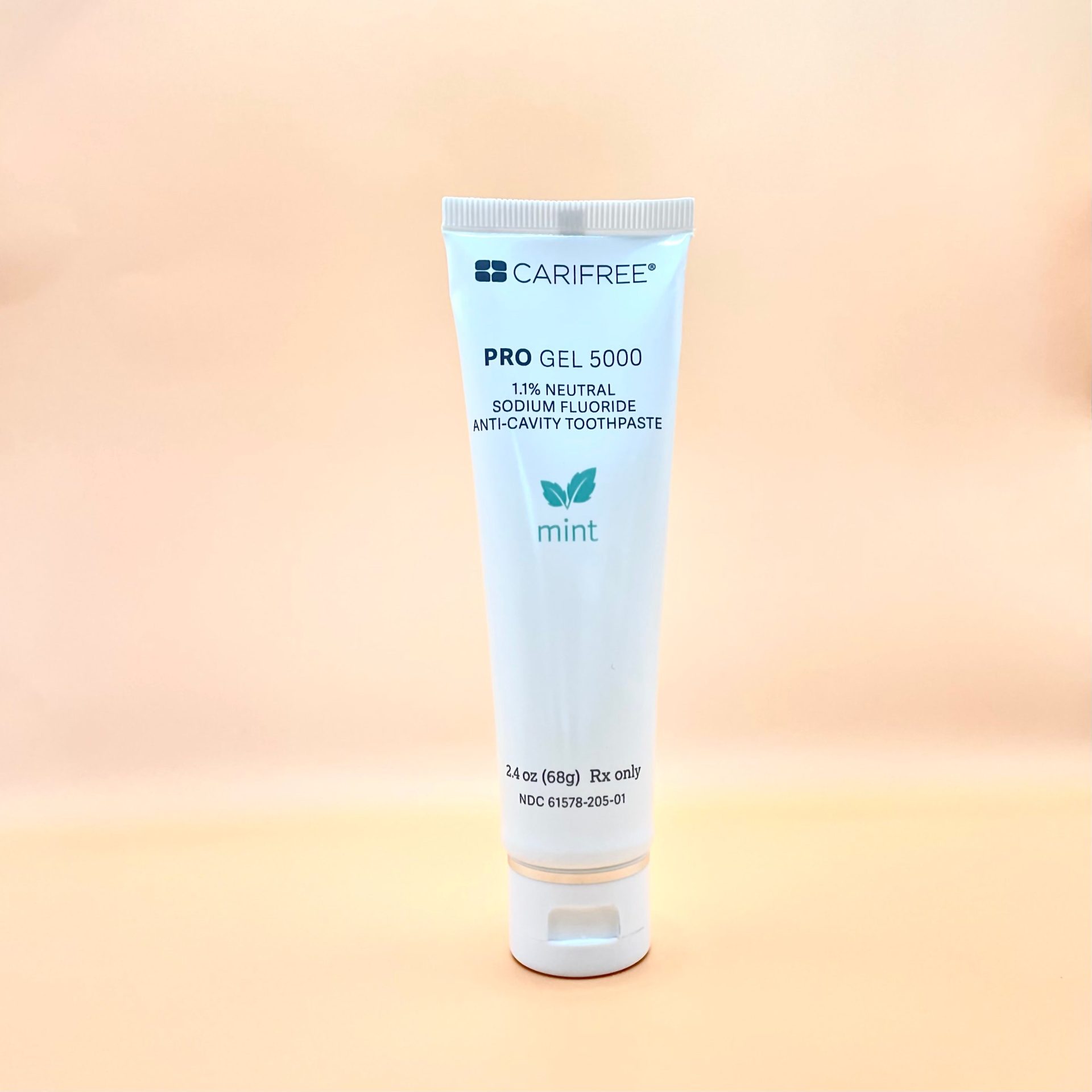
CariFree Pro Gel 5000
Prescription 5,000 ppm fluoride gel with elevated pH and xylitol — ideal for reflux, acidic diets, dry mouth, or sensitive gums.
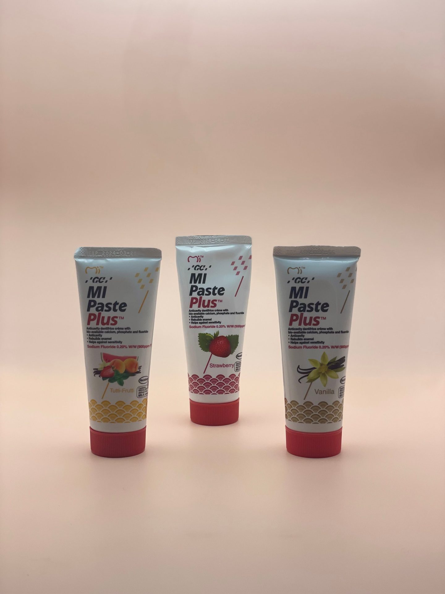
MI Paste Plus
Topical crème with CPP-ACP plus fluoride to remineralize enamel and relieve sensitivity.
About Dr. Sona Saeidi, DMD
Cofounder of Soothing Dental in San Francisco's Financial District. DMD magna cum laude, Boston University Goldman School of Dental Medicine. Practicing in California since 2011 (license #60566). Member of the American Dental Association, California Dental Association, and San Francisco Dental Society.
This page is educational and is not a diagnosis. Products sold through Soothing Dental may require a prescription; eligibility is confirmed through a brief telehealth screen at checkout.